
Introduction: The Architecture of Athletic Performance
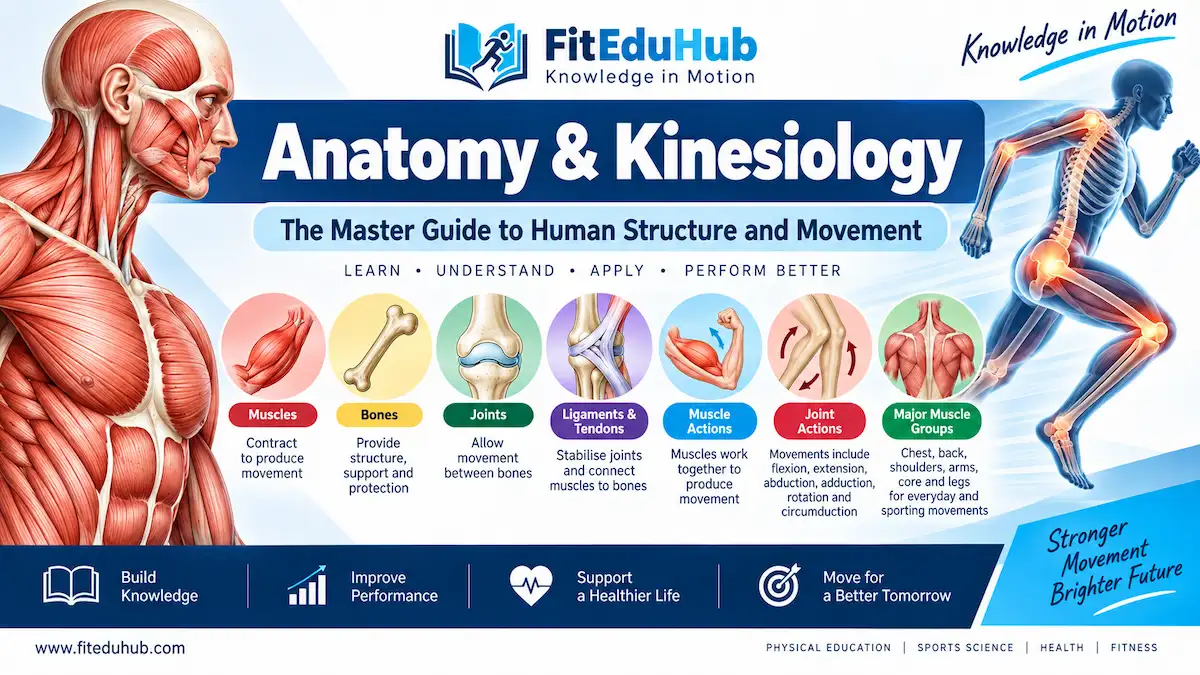
While sports biomechanics teaches us the invisible laws of physics (force, torque, and gravity), Anatomy and Kinesiology show us the physical machine that actually performs the work. Anatomy is the scientific study of the body’s physical structure, while kinesiology is the study of how those structures interact to create human movement.
To truly understand athletic performance, injury prevention, and physical rehabilitation, one must first understand the blueprint of the human body. Every sprint, jump, and lift relies on a perfectly synchronized symphony of bones acting as levers, joints acting as pivots, ligaments providing stability, and muscles firing as engines. In this comprehensive master guide, we will dissect the human musculoskeletal system and explore how anatomical structures dictate movement.
1. Bones: The Structural Framework (The Skeletal System)
The adult human body consists of 206 bones, which together form the skeletal system. In the context of sports science, bones do much more than simply give the body its shape. They are the foundational levers that muscles pull against to create movement.
Functions of Bones in Athletics
- Mechanical Leverage: Bones act as rigid levers. Without them, muscle contractions would simply cause the body to fold onto itself.
- Protection of Vital Organs: The skull protects the brain (crucial in contact sports like boxing), and the rib cage protects the heart and lungs.
- Mineral Storage & Blood Cell Production: Bones store calcium and phosphorus, and the bone marrow produces red blood cells (erythrocytes), which are essential for transporting oxygen to working muscles during endurance events like marathon running.
Types of Bones and Their Sports Applications
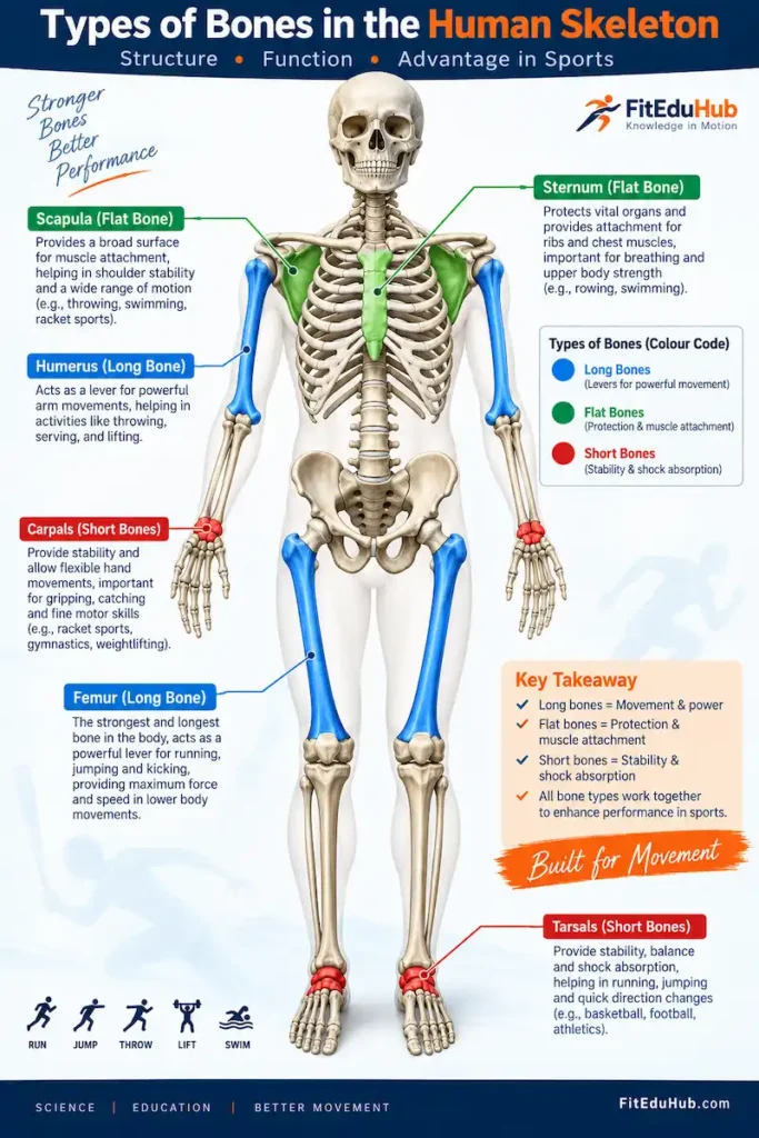
- Long Bones (e.g., Femur, Humerus, Tibia): These are longer than they are wide. They act as massive levers to generate speed and power.
- Practical Scientific Example: A taller volleyball player has a longer femur and tibia, giving them a longer biological lever to generate explosive vertical jumping power.
- Short Bones (e.g., Carpals in the wrist, Tarsals in the foot): Cube-shaped bones that provide stability and absorb heavy impacts, such as when a gymnast lands a dismount on the mat.
- Flat Bones (e.g., Scapula/Shoulder blade, Sternum): Provide broad surfaces for massive muscle attachments. The scapula is the anchor point for the muscles that control throwing velocity in a baseball pitcher.
- Irregular Bones (e.g., Vertebrae): Protect the spinal cord and allow for multi-directional flexibility of the torso.
👉 Pro Tip: Sports Biomechanics Learn exactly how these bone lengths affect force and torque in our Biomechanics guide on Levers.
2. Joints: The Hubs of Movement (The Articular System)
A joint (or articulation) is the exact location where two or more bones meet. While bones provide the rigid levers, joints provide the axes of rotation.
Classification of Joints
In kinesiology, we classify joints based on how much movement they allow:
- Fibrous Joints (Immovable): Held together by tough fibrous tissue. Example: The sutures in the skull. (Not highly relevant to movement).
- Cartilaginous Joints (Slightly Movable): Bones connected by cartilage. Example: The intervertebral discs in the spine, which compress slightly to absorb shock during heavy squats.
- Synovial Joints (Freely Movable): The most important joints in sports science. They contain a joint capsule filled with synovial fluid, which lubricates the joint to prevent friction during high-speed movements.
Key Synovial Joints in Human Movement
- Hinge Joints (e.g., Elbow, Knee): Allow movement in only one plane (Sagittal plane) – flexion and extension.
- Practical Scientific Example: The repetitive bending and straightening of the knee joint during cycling.
- Ball-and-Socket Joints (e.g., Shoulder, Hip): The most mobile joints in the body. The spherical head of one bone fits into the cup-like socket of another, allowing movement in all three planes (multi-axial).
- Practical Scientific Example: A swimmer performing the butterfly stroke relies on the immense rotational mobility of the shoulder’s ball-and-socket joint.
- Pivot Joints (e.g., Radioulnar joint in the forearm): Allows rotational movement around a single axis. Crucial for turning a doorknob or putting top-spin on a tennis forehand.
👉 Pro Tip: Sports Injury & Rehabilitation Discover how to treat and rehabilitate common joint injuries like shoulder dislocations in our Injury Management section.
3. Ligaments & Tendons: The Connective Network
To ensure that bones and joints do not simply fall apart under the immense stress of athletic competition, the body relies on two critical types of connective tissue: Ligaments and Tendons. It is a common mistake for beginners to confuse the two.
Ligaments (Bone to Bone)
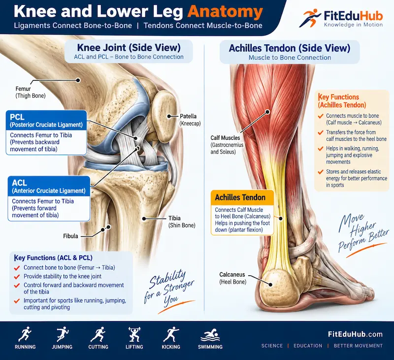
Ligaments are strong, elastic bands of connective tissue that connect bone to bone. Their primary job is to provide joint stability and prevent bones from moving outside their safe anatomical range.
- Tensile Strength: Ligaments are highly resistant to pulling forces but have very poor blood supply, which is why they heal very slowly when torn.
- Practical Scientific Example: The Anterior Cruciate Ligament (ACL) connects the femur (thigh bone) to the tibia (shin bone) inside the knee. When a football player abruptly plants their foot and twists their body to change direction, the ACL is the primary structure stopping the knee joint from collapsing or separating.
Tendons (Muscle to Bone)
Tendons are thick, cord-like tissues that connect muscle to bone. Their primary function is to transmit the mechanical force generated by a contracting muscle directly into the bone to create movement.
- The Stretch-Shortening Cycle (Elasticity): Tendons act like heavy-duty rubber bands. During athletic movements, they can stretch and store elastic energy, releasing it explosively.
- Practical Scientific Example: The Achilles tendon connects the massive calf muscles (gastrocnemius and soleus) to the heel bone. When a basketball player drops down to jump, the Achilles tendon stretches, stores kinetic energy, and then violently recoils, adding inches to their vertical leap without requiring extra muscle energy.
4. Muscles: The Engines of Movement (The Muscular System)
While bones, joints, and ligaments provide the physical hardware, skeletal muscles are the biological engines that power the human machine. There are over 600 skeletal muscles in the human body, making up about 40% of our total body weight.
The Structure of Skeletal Muscle
Skeletal muscle is highly organized and structured in bundles:
- Epimysium: The outer connective tissue wrapping the whole muscle.
- Fascicles: Bundles of individual muscle fibers within the muscle.
- Muscle Fibers (Myocytes): The actual muscle cells. They are cylindrical, multi-nucleated, and packed with mitochondria.
- Myofibrils & Sarcomeres: The microscopic contractile units of the muscle containing the proteins Actin (thin filaments) and Myosin (thick filaments).
How Muscles Contract: The Sliding Filament Theory
When your brain sends a neural signal to a muscle, calcium is released inside the muscle fiber. This allows the thick myosin filaments to grab onto the thin actin filaments and pull them toward the center. This microscopic sliding action shortens the entire muscle—this is what we call a muscle contraction.
👉 Pro Tip: Exercise Physiology To understand how ATP energy fuels this sliding filament contraction, read our deep dive into Exercise Energy Systems.
Muscle Fiber Types: The Genetics of Speed vs. Endurance
Not all muscle fibers are built the same. Kinesiologists classify them into two main categories:
- Type I (Slow-Twitch): Highly fatigue-resistant, dense with capillaries, and rely on oxygen. Perfect for endurance athletes like marathon runners.
- Type II (Fast-Twitch): Contract with explosive force and speed but fatigue very quickly. Perfect for powerlifters, Olympic weightlifters, and 100-meter sprinters.
5. Muscle Actions: The Physics of Muscle Contraction
Muscles do not simply turn “on” and “off.” Depending on the movement, they contract in completely different ways to manage force, generate speed, or absorb impact. In kinesiology, muscle contractions are classified into three main types.
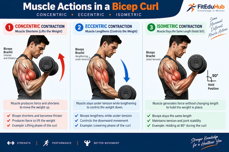
Concentric Contractions (Shortening)
- How it Works: The muscle produces enough force to overcome the external resistance (like gravity or a dumbbell), causing the muscle to shorten as it contracts.
- Practical Scientific Example: During the upward phase of a bicep curl, the bicep muscle contracts concentrically to lift the weight toward the shoulder. This is the acceleration phase of a movement.
Eccentric Contractions (Lengthening)
- How it Works: The muscle produces force, but it is less than the external resistance. As a result, the muscle lengthens while under tension.
- Practical Scientific Example: When you slowly lower the dumbbell back down during a bicep curl. The bicep is still working hard, acting as a biological brake to prevent the weight from simply dropping to the floor.
- Note: Eccentric contractions cause the most microscopic muscle damage, which is why they are primarily responsible for Delayed Onset Muscle Soreness (DOMS).
Isometric Contractions (Static)
- How it Works: The muscle generates internal force, but the length of the muscle does not change because the internal force exactly matches the external resistance. There is zero joint movement.
- Practical Scientific Example: Holding a plank position or pushing against an immovable wall. The muscles are firing maximally, but no physical movement occurs.
👉 Pro Tip: Recovery & Performance Learn how to recover faster from the heavy muscle damage caused by eccentric training in our DOMS & Recovery section.
6. Joint Actions: Roles of Muscles in Movement
A single muscle rarely works alone. To create smooth, coordinated human movement, muscles operate in highly synchronized teams. Kinesiologists define the roles of these muscles as follows:
The Agonist (Prime Mover)
The agonist is the main muscle responsible for producing a specific movement. It is the star of the show.
- Example: In a Bench Press, the Pectoralis Major (chest) is the agonist.
The Antagonist (The Opposer)
The antagonist is the muscle located on the exact opposite side of the joint from the agonist. It must relax and stretch to allow the agonist to contract. If it doesn’t, movement cannot occur.
- Example: In a Bicep Curl, the Triceps is the antagonist. It must relax so the bicep can shorten.
- Scientific Concept: This neurological process is called Reciprocal Inhibition. When your brain tells the agonist to fire, it simultaneously sends an inhibitory signal to the antagonist telling it to turn off.
The Synergists (The Assistants)
Synergists are smaller muscles that help the agonist perform the movement or guide the trajectory of the bone.
- Example: In a Bench Press, the Anterior Deltoid (front shoulder) and Triceps act as synergists to assist the chest.
The Stabilizers (The Fixators)
Stabilizers do not create the primary movement; instead, they contract isometrically to hold a joint or bone rigidly in place so the agonist has a firm base to pull from.
- Example: When doing a heavy Barbell Squat, the core muscles (Transverse Abdominis) and spinal erectors act as massive stabilizers to keep the spine rigid and safe.
👉 Pro Tip: Sports Biomechanics Understand how these synergist and stabilizer muscles manipulate the Centre of Gravity in our Biomechanics module.
7. Major Muscle Groups: The Athletic Engine Room
To design effective training programs or analyze sports movements, coaches must understand the major muscle groups and their primary anatomical functions.
The Upper Body
- Pectoralis Major (Chest): Responsible for horizontal adduction (bringing the arms across the chest). Crucial for throwing, punching, and pushing.
- Latissimus Dorsi (Lats): The widest muscle in the body. Responsible for shoulder extension and adduction (pulling the arms down and back). The powerhouse muscle for swimmers and rock climbers.
- Deltoids (Shoulders): Composed of three heads (Anterior, Lateral, Posterior). Responsible for raising the arms in all planes. Essential for racket sports and throwing.
- Biceps & Triceps (Arms): The biceps flex the elbow (pulling); the triceps extend the elbow (pushing).
The Core
- Rectus Abdominis: The “six-pack” muscle. Responsible for spinal flexion (crunching forward).
- Transverse Abdominis: The deepest core muscle. It acts like a biological weightlifting belt, wrapping around the spine to create intra-abdominal pressure and extreme stability.
The Lower Body (The Power Center)
- Gluteus Maximus: The largest and most powerful muscle in the human body. Responsible for hip extension. It is the primary engine for sprinting, jumping, and heavy squatting.
- Quadriceps: A group of four muscles on the front of the thigh. Responsible for knee extension (straightening the leg). Crucial for kicking and absorbing landing forces.
- Hamstrings: A group of three muscles on the back of the thigh. Responsible for knee flexion and assisting in hip extension. They are the most commonly torn muscles in sprinting.
- Gastrocnemius & Soleus (Calves): Responsible for plantarflexion (pointing the toes). Essential for acceleration and explosive jumping.
👉 Pro Tip: Strength & Conditioning Learn how to safely overload and build strength in all these major muscle groups in our Resistance Training guide.
Conclusion: Mastering the Human Machine
Anatomy and Kinesiology are not just topics for medical students; they are the fundamental alphabets of athletic coaching and performance. By understanding which bones act as levers, how the synovial joints pivot, and exactly how the agonists and antagonists interact, we move away from guessing and step into the realm of precise, scientific training.
Whether your goal is to rehab a torn ligament, increase sprinting speed, or design a flawless hypertrophy program, mastering the anatomical structure of the human body is your ultimate blueprint for success.
📚 Evidence-Based References
To continue your deep dive into the anatomy of human movement, we recommend these two highly respected foundational texts:
- Manual of Structural Kinesiology by R.T. Floyd. (The ultimate standard text for understanding the specific origins, insertions, and actions of every major skeletal muscle).
- Trail Guide to the Body by Andrew Biel. (An exceptional, highly visual resource for locating and understanding the practical function of bones, joints, and ligaments).