

Introduction: The Unseen Phase of Athletic Growth
There is a pervasive misconception across sports science and strength conditioning that athletes build strength, speed, and cardiovascular power during their workouts. From an exercise physiology perspective, this is fundamentally false. Physical training is merely a mechanical and metabolic stimulus designed to disrupt physiological homeostasis, induce microtrauma in skeletal muscle fibers, and deplete cellular energy reserves.
The actual biological adaptation, cellular reconstruction, and functional growth occur exclusively during Recovery.
Without a systematically structured recovery strategy, an athlete continuously accumulates residual physiological strain, eventually leading to non-functional overreaching or severe overtraining. High-performance optimization relies on balancing training stress with biological repair mechanisms. This master guide examines the cellular processes of muscle regeneration, the hormonal cascades of sleep, fatigue mechanics, and workload quantification methods used in elite sports.
Athletic Recovery & Readiness Dashboard
Physiological Status: Optimal1. Rest vs. Recovery: The Biological Imperative
While “rest” and “recovery” are often used interchangeably, sports scientists apply strict physiological distinctions between the two processes.
PASSIVE REST (Cessation)
- CNS Power Down & Neural Reset
- Baseline Cardiovascular Restoration
- Passive Sleep Architecture
ACTIVE RECOVERY (Strategic Modality)
- Metabolic Waste & H+ Clearance
- Sub-Maximal Local Vasodilation
- Targeted Nutrient Delivery
Passive Rest
Passive rest denotes the complete cessation of movement and physical exertion. It allows the Central Nervous System (CNS) to reduce motor unit firing frequencies and permits the cardiovascular system to return to a resting baseline. Sleep and static relaxation represent primary forms of passive rest. While essential, passive rest alone is insufficient for rapid metabolic waste clearance following high-intensity training.
Active Recovery
Active recovery encompasses deliberate, sub-maximal activities designed to accelerate biological tissue repair and restore physiological homeostasis. Key facets include:
- Nutritional Recovery: Systematic replenishment of muscle glycogen stores and provision of essential amino acids to initiate protein synthesis.
- Active Hyperaemia: Engaging in low-impact, low-intensity exercise (e.g., 20 minutes at under 50% VO2 max) to promote muscular blood flow, delivering oxygen while clearing hydrogen ions and inorganic phosphate.
- Psychological Recovery: Mental detachment from competitive stressors to decrease tonic sympathetic nervous system activity.
PRO TIP: EXERCISE PHYSIOLOGY INTEGRATION
Understanding how the human body converts macronutrients into adenosine triphosphate (ATP) during recovery is vital for athletic performance. Learn more in our comprehensiveExercise Physiology Guide.
2. Sleep: The Ultimate Performance Enhancer
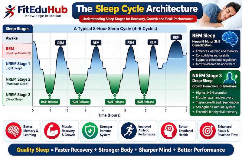
Sleep represents the most critical, evidence-based recovery modality in human physiology. Cognitive or physical interventions cannot replace the biological adaptations occurring during structured sleep cycles.
NON-REM (SLOW-WAVE DEEP SLEEP)
- Anabolic Somatotropin / HGH Peak Release
- Accelerated Systemic Muscle Protein Synthesis
- Structural Myofibrillar Microtrauma Repair
- Immune System Cytokine Secretion
REM (RAPID EYE MOVEMENT) SLEEP
- Motor Skill Memory & Technical Consolidation
- Central Nervous System Neurotransmitter Reset
- Psychological & Cognitive Stress Recovery
- Neural Pathway Plasticity Reinforcement
Slow-Wave Sleep (Non-REM) and Anabolic Hormonal Cascades
During Stage 3 Non-Rapid Eye Movement (NREM) sleep—also termed Slow-Wave Sleep (SWS)—the body enters its primary anabolic state:
- Human Growth Hormone (HGH) Secretion: The anterior pituitary gland releases large pulses of somatotropin (HGH). This hormone stimulates cellular amino acid uptake, enhances tissue repair, and promotes systemic protein synthesis.
- Testosterone Production: Peak systemic testosterone secretion occurs during undisturbed sleep cycles, supporting myofibrillar growth and central nervous system recovery.
- Cortisol Dynamics: Sleep deprivation disrupts the diurnal cortisol rhythm, causing sustained elevations in circulating glucocorticoids. Chronically elevated cortisol accelerates muscle protein breakdown and inhibits anabolic signaling pathways.
REM Sleep and Motor Learning Consolidation
Rapid Eye Movement (REM) sleep facilitates neurological recovery. During REM sleep, the brain consolidates motor patterns acquired during training sessions, translating technical skill practice into permanent neural adaptations.
3. Muscle Recovery: The Cellular Reconstruction Process
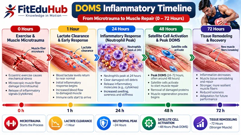
High-intensity resistance training or rapid decelerations induce microscopic disruptions within the structural architecture of skeletal muscle fibers, specifically damaging the Z-discs and contractile proteins (actin and myosin).
The Cellular Regeneration Pipeline
- Inflammatory Phase: Immediately following mechanical trauma, neutrophils and macrophages infiltrate the damaged tissue. Macrophages clean cellular debris and release pro-inflammatory cytokines, initiating tissue reconstruction.
- Satellite Cell Activation: Satellite cells—quiescent myogenic stem cells located between the basal lamina and sarcolemma—activate in response to mechanical strain.
- Proliferation and Fusion: Activated satellite cells proliferate, differentiate, and fuse with damaged muscle fibers or form new myotubes. This process donates new myonuclei to the damaged fibers, expanding their transcriptional capacity for protein synthesis and hypertrophy.
PRO TIP: ANATOMY & KINESIOLOGY INTEGRATION
To understand how microscopic muscle tearing affects sliding filament mechanics and muscle cross-sectional area, review ourAnatomy & Kinesiology Master Guide.
4. DOMS (Delayed Onset Muscle Soreness) Mechanics
Delayed Onset Muscle Soreness (DOMS) represents a distinct physiological phenomenon characterized by muscular stiffness, localized tenderness, and reduced force development, typically peaking 24 to 72 hours post-exercise.
| Parameter | The Lactic Acid Myth | The Scientific Reality of DOMS |
| Primary Cause | Supposed accumulation of metabolic lactate in muscle tissue | Microscopic structural Z-disc disruption and inflammatory cascade |
| Clearance Timeline | Blood lactate returns to baseline within 30–60 minutes post-exercise | Inflammatory response peaks at 48 hours post-stimulus |
| Contraction Type | Incorrectly linked to any high-intensity activity | Primarily induced by high-force eccentric lengthenings |
| Primary Treatment | Static resting or passive stretching | Sub-maximal active recovery promoting local hyperaemia |
The Eccentric Mechanical Driver
DOMS is primarily caused by unaccustomed eccentric contractions—where muscles produce force while lengthening (e.g., the lowering phase of a heavy squat or downhill running). High force per unit of muscle cross-sectional area during eccentric actions creates mechanical strain on structural proteins.
Evidence-Based Management of DOMS
- Avoid Over-Reliance on NSAIDs: Non-Steroidal Anti-Inflammatory Drugs (e.g., ibuprofen) reduce acute soreness, but research demonstrates they blunt inflammatory signaling pathways required for satellite cell activation, potentially compromising long-term adaptations.
- Active Hyperaemia: Low-intensity exercise increases localized microvascular perfusion, accelerating the removal of inflammatory cellular byproducts without blunting adaptation.
5. Fatigue Mechanics: Peripheral vs. Central Fatigue
Fatigue represents a protective biological mechanism regulated by peripheral tissue environments and the Central Nervous System (CNS) to prevent catastrophic physiological damage.
PERIPHERAL FATIGUE (Muscular)
- Intramuscular Acidosis (H+ Accumulation)
- Inorganic Phosphate (Pi) Accumulation
- Impaired Sarcoplasmic Ca2+ Release
- Disrupted Cross-Bridge Cycling Mechanics
CENTRAL FATIGUE (Nervous System)
- Group III & IV Inhibitory Afferent Signaling
- Suppressed Motor Cortex Descending Output
- Central Neurotransmitter Imbalances
- Protective Neural Brake Activation
Peripheral Fatigue (Muscular Level)
Peripheral fatigue occurs distal to the neuromuscular junction within the skeletal muscle fibers:
- Intramuscular Acidosis: Rapid accumulation of hydrogen ions during high-intensity glycolytic exercise lowers intramuscular pH.
- Excitation-Contraction Uncoupling: Elevated inorganic phosphate levels interfere with calcium ion release from the sarcoplasmic reticulum, directly reducing cross-bridge cycling force output.
Central Fatigue (Nervous System Level)
Central fatigue originates within the central nervous system (brain and spinal cord):
- Decreased Motor Drive: The brain decreases the amplitude and frequency of descending motor signals sent to working muscle groups.
- Afferent Feedback: Group III and IV muscle afferent nerves detect metabolic stress, tissue damage, and elevated core body temperature, sending inhibitory signals to the motor cortex to suppress motor output.
PRO TIP: SPORTS BIOMECHANICS INTEGRATION
Central and peripheral fatigue directly alter joint kinematics, ground reaction forces, and movement efficiency. Explore force-velocity profiling in ourSports Biomechanics Master Guide.
6. Training Load Quantification: The ACWR Framework
Effective recovery management relies on objective quantification of training stress.
Acute:Chronic Workload Ratio (ACWR) Safety Spectrum
Internal vs. External Load
- External Load: Objective measures of work performed (e.g., total volume load in kilograms, sprint distance in meters, or mechanical power output in watts).
- Internal Load: Physiological and psychological stress elicited by the external load (e.g., Heart Rate Variability, session RPE, or blood lactate concentrations).
The Acute:Chronic Workload Ratio (ACWR) Spectrum
- Under-Training Zone (Below 0.8): Fitness decays, increasing relative injury risk upon re-exposure to intense workloads.
- Optimal “Sweet Spot” (0.8 to 1.3): Balances fitness adaptations while keeping injury risk low.
- Danger Zone (Above 1.5): Rapid workload spikes significantly increase injury risk and central fatigue accumulation.
PRO TIP: SPORTS RESEARCH & TECHNOLOGY INTEGRATION
Advanced GPS micro-pods, velocity-based transducers, and force plate telemetry streamline ACWR monitoring. Learn more in ourSports Research & Technology Guide.
7. Overtraining Syndrome (OTS): Clinical Breakdown
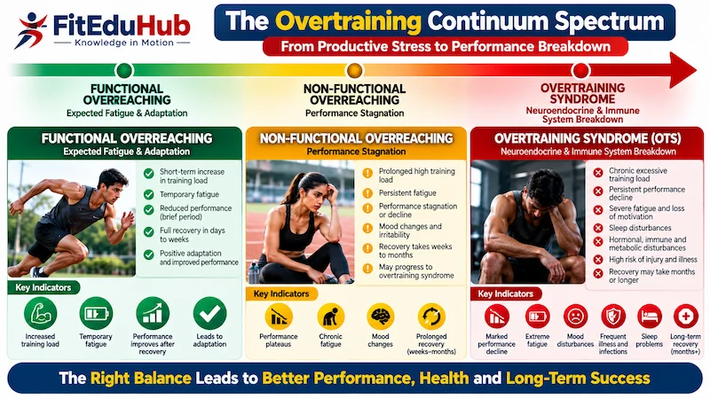
When an athlete sustains excessive training volumes or intensities without adequate recovery over weeks or months, they risk developing Overtraining Syndrome (OTS)—a complex neuroendocrine disorder.
THE OVERTRAINING CONTINUUM
Functional Overreaching
Short-term fatigue resulting in supercompensation and improved performance following planned deloads.
Non-Functional Overreaching
Stagnant or declining performance requiring weeks of rest to fully restore baseline capacities.
Overtraining Syndrome (OTS)
Systemic neuroendocrine and immune breakdown requiring months of clinical recovery.
Key Clinical Indicators of OTS
- Autonomic Dysregulation: Persistent changes in resting heart rate (over 10 bpm elevation) and blunted heart rate recovery following standardized sub-maximal tests.
- Neuroendocrine Dysfunction: Suppression of the hypothalamic-pituitary-adrenal (HPA) axis, resulting in flattened cortisol rhythms and reduced circulating testosterone-to-cortisol ratios.
- Immunological Depression: Increased susceptibility to upper respiratory tract infections (URTIs) driven by decreased mucosal IgA levels.
PRO TIP: STRENGTH & CONDITIONING INTEGRATION
Preventing OTS requires structured periodization and planned deload weeks. Master periodization strategies in ourStrength & Conditioning Guide.
8. Evidence-Based Recovery Interventions
| Recovery Modality | Primary Biological Mechanism | Optimal Application Window | Key Scientific Caveat |
| Cold Water Immersion (CWI) | Acute vasoconstriction followed by hyperaemia; reduces tissue temperature and microvascular permeability | Multi-day tournament settings requiring rapid turnaround (under 24 hours) | Blunts local inflammatory signaling required for long-term muscle hypertrophy |
| Self-Myofascial Release (Foam Rolling) | Reduces arterial stiffness, enhances local vascular endothelial function, and attenuates DOMS perception | Post-training or during active recovery days | Does not physically lengthen structural fascia permanently |
| Pneumatic Compression Boots | Enhanced venous return and lymphatic drainage of metabolic byproducts | Post-competition or during travel | Primarily impacts acute subjective recovery perception |
Conclusion: The Ultimate Balancing Act
In high-performance sports, physical training sets the potential for adaptation, but recovery dictates whether that potential is realized. Athletes who train rigorously without equal commitment to recovery fail to reach peak performance.
Optimizing recovery requires balancing physiological stress and active repair: prioritizing deep sleep architecture, managing muscle microtrauma, tracking ACWR metrics, and applying targeted recovery modalities. Managing this balance allows athletes to maximize adaptation while minimizing injury risk.
Academic References & Peer-Reviewed Literature
- 1. Fullagar, H. H., et al. (2015). Sleep and athletic performance: the effects of sleep loss on exercise performance and physiological function. Sports Medicine, 45(2), 161-186. [PubMed Citation]
- 2. Gabbett, T. J. (2016). The training—injury prevention paradox: should athletes be training smarter and harder? British Journal of Sports Medicine, 50(5), 273-280. [BJSM Citation]
- 3. Peake, J. M., et al. (2017). Muscle damage and inflammation during recovery from exercise. Journal of Applied Physiology, 122(3), 559-570. [PubMed Citation]
- 4. Meeusen, R., et al. (2013). Prevention, diagnosis, and treatment of the overtraining syndrome: joint consensus statement of the ECSS and ACSM. Medicine & Science in Sports & Exercise, 45(1), 186-205. [ACSM Citation]
Frequently Asked Questions: Recovery & Performance
Answer: Physical training serves purely as a mechanical and metabolic stressor that disrupts cellular homeostasis and creates muscle microtrauma. Supercompensation, myofibrillar tissue reconstruction, and cellular growth occur exclusively during rest and recovery phases when anabolic hormones peak.
Answer: During Slow-Wave Sleep, the anterior pituitary gland releases major pulses of Human Growth Hormone (HGH). HGH stimulates cellular amino acid uptake and protein synthesis, restoring micro-damaged structural myofibrils.
Answer: Satellite cells are myogenic stem cells located on the exterior of muscle fibers. Exercise-induced mechanical strain activates satellite cells, causing them to proliferate and fuse with damaged myofibers, donating new myonuclei to expand protein synthesis capacity.
Answer: Blood lactate is fully cleared from muscle tissue within 30 to 60 minutes post-exercise. DOMS is caused by microscopic Z-disc disruption and localized inflammatory signaling cascades that peak 24 to 48 hours later, particularly following eccentric contractions.
Answer: Peripheral fatigue occurs within the muscle tissue due to metabolic byproduct accumulation (H+ and Pi) and impaired calcium release. Central fatigue originates in the central nervous system, where the brain reduces descending motor drive to protect organ systems from excessive stress.
Answer: ACWR divides acute training load (7-day sum) by chronic training capacity (28-day rolling average). The optimal “sweet spot” is 0.8 to 1.3, maintaining adaptation while minimizing injury risk. Values exceeding 1.5 significantly increase injury likelihood.
Answer: NSAIDs inhibit COX enzymes and suppress the acute inflammatory signaling required to activate satellite cells. While they reduce acute DOMS, long-term use can blunt myofibrillar hypertrophy and structural adaptation.
Answer: Primary indicators include autonomic nervous system dysregulation (elevated resting heart rate), blunted cortisol responses due to HPA-axis exhaustion, reduced testosterone-to-cortisol ratios, and decreased mucosal IgA levels causing frequent upper respiratory infections.
Answer: CWI should be avoided immediately following hypertrophy or maximal strength resistance training sessions. The severe vasoconstriction and acute temperature reduction blunt localized inflammatory signaling required for long-term anabolic adaptations.
Answer: Active recovery promotes low-level hyperaemia without causing mechanical damage or central fatigue. Increased muscular blood flow speeds the removal of cellular metabolic byproducts (H+ and Pi) and delivers oxygen and nutrients to recovering tissue.
Sports Recovery and Training Adaptation